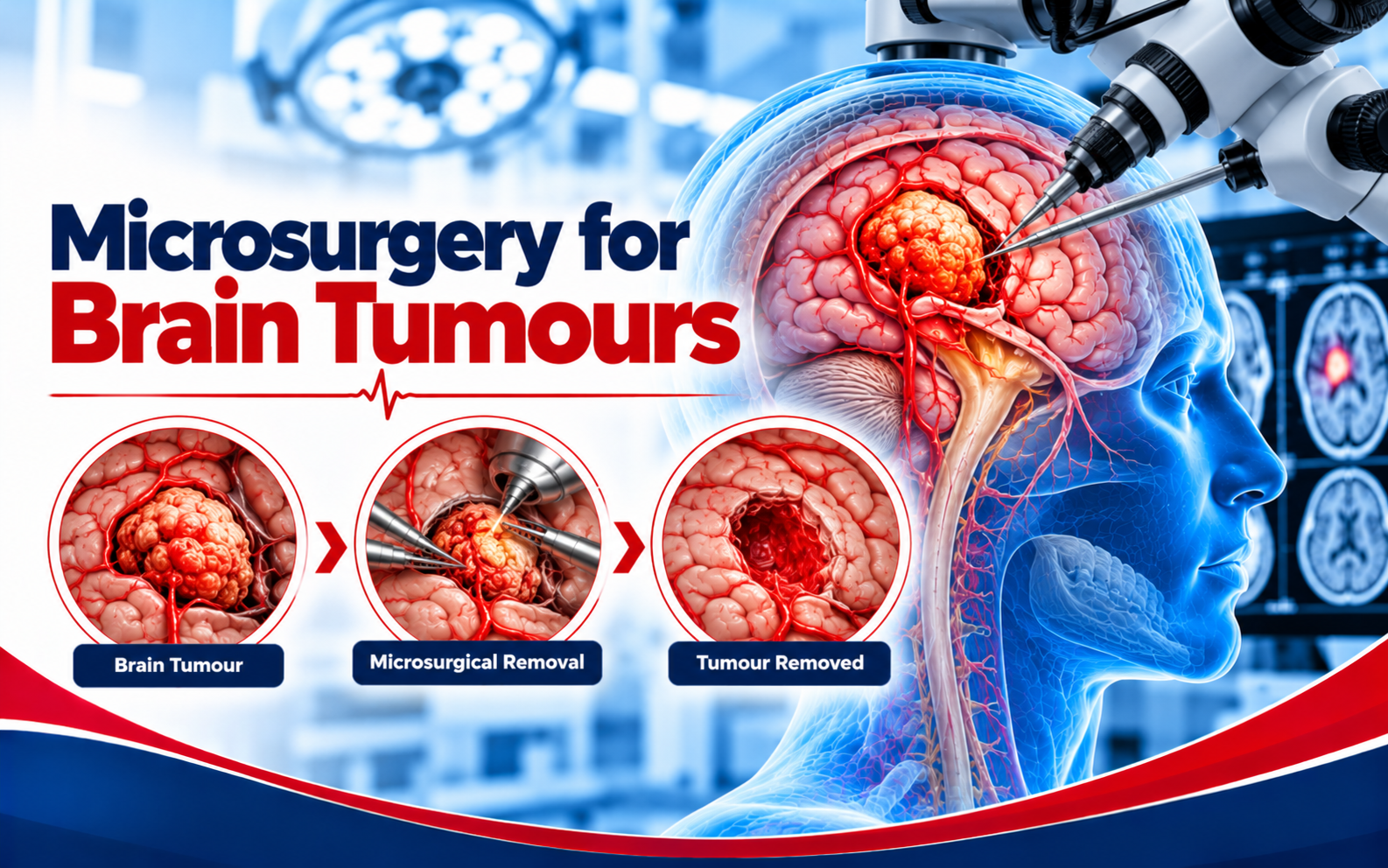
Microsurgical planning for selected brain tumours.
Understanding Microsurgery for Brain Tumours and the reasons for specialist review
Brain tumour microsurgery uses operating microscopes and careful planning to access and treat selected intracranial tumours. The correct first step is to understand the diagnosis rather than to select an operation from a website. Different people can have similar symptoms but different scan findings, and even the same scan result can mean different things depending on a person’s examination and clinical history. A careful discussion brings symptoms, physical findings, images and individual priorities together before treatment is recommended.
For someone looking into microsurgery for brain tumours in Noida, it helps to know that this page explains the general medical decision-making process; it does not establish that any specific procedure will be available or appropriate. The reason for a consultation may be a new finding on imaging, ongoing symptoms, changing function, or the need to understand a recommendation already given elsewhere. Each of these starting points calls for a different conversation.
Symptoms and situations that deserve attention
People with an imaging-detected brain lesion, seizures, persistent neurological symptoms or a need for a surgical opinion may benefit from specialist consultation. Symptoms in this area may include a newly developed seizure, persistent change in strength or speech, progressive headache, visual disturbance or other neurological symptoms. Their meaning depends on the pattern, onset, severity and associated findings. For example, a sudden change may have a very different urgency from a symptom that has been stable for months. It is useful to note the first day something changed and whether it is improving, fluctuating or steadily worsening.
Changes in concentration, speech, mood, work and family responsibilities may be relevant to decisions and recovery planning. Do not assume that the intensity of pain or discomfort reliably measures the seriousness of a condition. Some important neurological problems affect strength, balance, sensation or coordination more than they cause pain. A structured symptom history can help the treating team focus an examination and decide whether emergency or planned care is required.
How a diagnosis is assessed
MRI findings, tumour position, symptoms and overall medical condition are reviewed, sometimes with input from other specialists. Depending on the presenting concern, assessment may involve neurological examination, MRI and CT where appropriate, and further specialist imaging or tests when needed. The chosen investigations should answer a specific clinical question. More tests are not always better, and repeating an image without a clear reason may not help. Clinicians also consider relevant medical conditions, current medicines, allergies, previous operations and the patient’s functional baseline.
When reviewing microsurgery for brain tumours, a specialist compares the radiology report with the actual images and with the examination. The written report is valuable, but it may not tell the entire story. A discussion should distinguish what is confirmed, what remains uncertain, and whether another specialist, an additional investigation or a period of observation would change the plan.
What imaging can and cannot tell you
Imaging provides a view of anatomy at a particular moment, whereas symptoms and neurological examination show how the person is functioning. An abnormality on a scan is not automatically the explanation for every symptom. Equally, a change in strength, walking, speech or sensation can require further assessment even when an initial report sounds reassuring. Both aspects are considered together, with attention to the timing of the scan and any change since it was performed.
For microsurgery for brain tumours, questions about size, exact position, surrounding structures and stability can be particularly relevant. Ask the clinician to point out the key finding and explain how confidently it relates to your symptoms. If an image was taken elsewhere, bringing the scan files rather than only a photograph of the report can make an in-person review more useful.
Treatment choices and their alternatives
Treatment may involve biopsy, resection, observation or nonsurgical care. Surgical aims depend on the tumour type and its relationship to critical brain structures. Possible pathways can also include careful observation, medication, biopsy, surgery, radiotherapy or other specialty-led treatment according to the diagnosis. The best option is not necessarily the most technically advanced one; it is the option whose expected benefit is justified by the person’s diagnosis, risks and goals. Some problems require urgent intervention, while others can reasonably be monitored or treated without an operation.
Before deciding on microsurgery for brain tumours, patients and families can ask what problem the proposed treatment is intended to solve, which symptoms might not improve, and what may happen if intervention is deferred. These questions help separate the treatment goal from unrealistic expectations. A balanced consultation discusses potential advantages and limitations rather than promising a guaranteed result.
What a procedure or treatment pathway may involve
If intervention is recommended, the treating hospital explains the planned approach, setting, anaesthesia, monitoring and likely sequence of care. The exact steps vary widely across brain, spine and blood-vessel procedures and may change when additional information becomes available. Patients should know which team is responsible at each stage, whether another specialist is involved, and how the plan could be adjusted if findings differ from expectations.
In a discussion about microsurgery for brain tumours, it is reasonable to ask why this specific route is preferred over a different technique. A smaller incision, catheter-based device or newer technology can have advantages in selected circumstances but is not automatically safer or better for everyone. The purpose of the technology should be explained in everyday language and linked to the individual clinical problem.
Benefits, uncertainties and possible complications
The hoped-for benefit of microsurgery for brain tumours must be weighed against its limitations. Depending on the procedure, risks may include bleeding, infection, neurological deficits, seizures, swelling and complications linked to the precise procedure. Risk is not identical for every patient; age, other illnesses, medicines and anatomical details may influence it. A surgical discussion should explain the most relevant possibilities, how complications would be recognised, and what support is available if recovery is more difficult than expected.
No responsible clinician can promise a particular result on the basis of a website description. The outcome may depend on the condition itself as well as the chosen treatment. The right goal can sometimes be to prevent deterioration or obtain a diagnosis rather than to remove every symptom. Asking about these distinctions can make consent more informed and reduce misunderstandings after care.
Preparing for an informed consultation
Bring a list of current medicines, including blood thinners and supplements, along with previous prescriptions, discharge summaries, available scan images, relevant laboratory reports and known allergies. A short timeline describing the first symptoms, their progression and previous treatments is often more helpful than a large number of unsorted documents. When possible, write down two or three important questions before the appointment.
For a microsurgery for brain tumours appointment, describe what you can no longer do comfortably, rather than relying only on a pain score or scan label. Mention falls, weakness, loss of independence, new seizures, changes in speech or difficulty with ordinary tasks when relevant. Family observations can add useful context, particularly when changes have been gradual or affect awareness and memory.
Recovery, rehabilitation and follow-up
Care does not stop when a procedure ends. Follow-up may include hospital neurological assessments, careful medication review, rehabilitation when required and planned imaging or pathology follow-up. Some patients need therapy or additional input from other medical specialists. The expected course varies, so the treating team should provide instructions tailored to the actual procedure and to any neurological changes present before treatment. Progress is assessed against realistic goals rather than a fixed recovery timetable taken from the internet.
Before leaving hospital or completing a specialist visit, clarify the purpose and timing of the next review, how medicines should be taken, which activities can be resumed, and which symptoms require urgent reassessment. Keeping reports and discharge paperwork organised can reduce delays if advice is needed later. A symptom that is new, progressive or very different from the original complaint deserves fresh medical attention.
When an emergency cannot wait
Diagnosis, prognosis and treatment options differ significantly between tumour types; individual counselling is essential. Warning signs for urgent assessment may include a first seizure lasting several minutes, new one-sided weakness, an abrupt severe headache or a significant reduction in consciousness. These are examples rather than a complete checklist. If a symptom suggests stroke, a serious brain bleed, acute spinal cord compression or a prolonged seizure, seek emergency services and an equipped hospital immediately instead of filling out an online appointment form.
Time can matter greatly in some neurological emergencies. Do not delay emergency assessment to contact a particular doctor or arrange a routine second opinion. A clinician at the receiving hospital can decide which investigations and specialists are needed as the situation evolves. Planned consultation is appropriate for stable questions, reports and follow-up discussions, not for rapidly changing neurological function.
Planning Microsurgery for Brain Tumours care in Noida
People searching for microsurgery for brain tumours in Noida may be trying to compare options, obtain a second opinion or understand a diagnosis made during a recent hospital visit. Ask about the consultation location, any available hospital facilities, the relevant care team and the practical arrangements for investigations and follow-up. It is sensible to confirm appointments directly before travelling because schedules and treatment availability may change.
Dr. Yashpal Singh Bundela’s supplied professional profile lists experience in neurosurgery and consultations in Noida. The information on this website is intended to support patient understanding, not to replace an assessment or imply that a procedure is guaranteed at a particular facility. A useful appointment should leave you with a clearer explanation of the problem and the reasons behind the proposed next step.
Questions you can ask your specialist
What do the images suggest, is more diagnostic testing needed, and how will the team protect important brain functions? Other useful questions include whether the condition is stable or progressing, which test findings are most important, and whether any change in symptoms would alter the urgency. If a procedure is being considered, ask about the specific goal, the main risks, hospital care, recovery support and alternative options. A written set of questions can help family members participate without losing track of the discussion.
For microsurgery for brain tumours, ask what successful management would mean in your own circumstances. It might be improved function, prevention of further harm, diagnostic clarity or control of a particular symptom. Understanding that target makes it easier to weigh benefits against burdens. If the explanation is confusing, request simpler language or ask for the relevant image to be reviewed again.
Frequently asked questions
Can all brain tumours be completely removed? No. The safe extent of surgery varies with the tumour’s location, type and surrounding brain structures. Decisions depend on current symptoms, examination findings, relevant investigations and a full discussion of alternatives. No single rule can replace an individual evaluation, and it can be appropriate to seek another specialist opinion when the situation is stable and important choices remain uncertain.
How soon should I seek advice about microsurgery for brain tumours? The answer depends on whether symptoms are sudden, progressive, disabling or stable. Red flags warrant emergency assessment, whereas nonurgent questions can usually begin with a scheduled clinical review. Bring the original investigations and ask the team what changes should prompt faster reassessment.
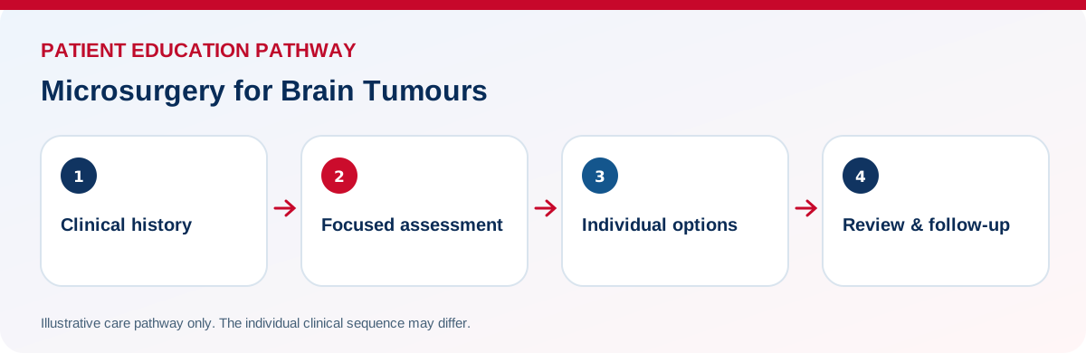
Discuss symptoms, imaging, treatment alternatives and expected follow-up.